Chapter 5
Tissues
This chapter was edited and adapted from chapter “The Tissue Level Organization” of the open source book Anatomy and Physiology 2e from OpenStax (original text available for free at https://openstax.org/details/books/anatomy-and-physiology-2e).

The body contains at least 200 distinct cell types. These cells contain essentially the same internal structures yet they vary enormously in shape and function. The different types of cells are not randomly distributed throughout the body; rather they occur in organized layers, a level of organization referred to as tissue. Figure 4.1 shows the high degree of organization among different types of cells in the tissue of the cervix. You can also see how that organization breaks down when cancer takes over the regular mitotic functioning of a cell (red arrows). The variety in shape reflects the many different roles that cells fulfill in your body. The human body starts as a single cell at fertilization. As this fertilized egg divides, it gives rise to trillions of cells, each built from the same blueprint, but organizing into tissues and becoming irreversibly committed to a developmental pathway.
The term tissue is used to describe a group of cells found together in the body. The cells within a tissue share a common embryonic origin. Microscopic observation reveals that the cells in a tissue share morphological features and are arranged in an orderly pattern that achieves the tissue’s functions. From the evolutionary perspective, tissues appear in more complex organisms. For example, multicellular protists, ancient eukaryotes, do not have cells organized into tissues.
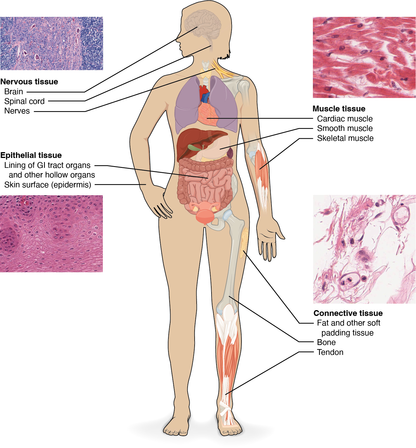
Although there are many types of cells in the human body, they are organized into four broad categories of tissues: epithelial, connective, muscle, and nervous (Figure 4.2). Each of these categories is characterized by specific functions that contribute to the overall health and maintenance of the body. A disruption of the structure is a sign of injury or disease. Such changes can be detected through \textbf{histology}, the microscopic study of tissue appearance, organization, and function.
This chapter introduces all four tissue types. It will discuss in more detail epithelial tissues. Connective, muscle, and nervous tissues will be discussed in more detail in later chapters.
