Chapter 5
Tissues
5.1 Epithelial Tissue
This section was edited and adapted from chapter 4.2 “The Tissue Level Organization: Epithelial Tissue” of the open source book Anatomy and Physiology 2e from OpenStax (original text available for free at https://openstax.org/details/books/anatomy-and-physiology-2e).
Objectives
By the end of this section, you will be able to:
-
Explain the structure and function of epithelial tissue
-
Distinguish between simple epithelia and stratified epithelia, as well as between squamous, cuboidal, and, columnar epithelia
-
Describe the structure and function of endocrine and exocrine glands and their respective secretions
Epithelial tissue, also referred to as epithelium (plural: epithelia), refers to the sheets of cells that cover exterior surfaces of the body, line internal cavities and passageways, and form certain glands.
Skin is not the only area of the body exposed to the outside. Other areas include the airways, the digestive tract, as well as the urinary and reproductive systems, all of which are lined by an epithelium. Hollow organs and body cavities that do not connect to the exterior of the body, which includes, blood vessels and serous membranes, are lined by endothelium (plural = endothelia), which is a type of epithelium.
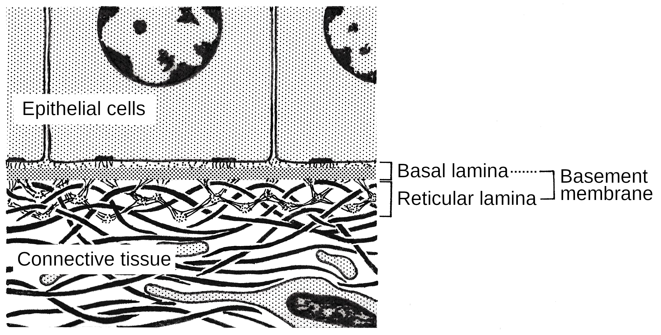
All epithelia share some important structural and functional features. This tissue is highly cellular, with little or no extracellular material present between cells. Adjoining cells form a specialized intercellular connection between their cell membranes called a cell junction. The epithelial cells exhibit polarity with differences in structure and function between the exposed or apical (top) facing surface of the cell and the basal (bottom) surface close to the underlying body structures. The basal lamina, a mixture of glycoproteins and collagen, provides an attachment site for the epithelium, separating it from underlying connective tissue. The basal lamina attaches to a reticular lamina, which is secreted by the underlying connective tissue, forming a basement membrane that helps hold it all together (Figure 5.3).
Epithelial tissues are nearly completely avascular (without blood vessels). For instance, no blood vessels cross the basement membrane to enter the tissue, and nutrients must come by diffusion or absorption from underlying tissues or the surface. Many epithelial tissues are capable of rapidly replacing damaged and dead cells. Sloughing off of damaged or dead cells is a characteristic of surface epithelium and allows our airways and digestive tracts to rapidly replace damaged cells with new cells.
Generalized Functions of Epithelial Tissue
Epithelial tissues provide the body’s first line of protection from physical, chemical, and biological wear and tear. The cells of an epithelium act as gatekeepers of the body controlling permeability and allowing selective transfer of materials across a physical barrier. All substances that enter the body must cross an epithelium. Some epithelia often include structural features that allow the selective transport of molecules and ions across their cell membranes. Many epithelial cells are capable of secretion and release mucous and specific chemical compounds onto their apical surfaces. The epithelium of the small intestine releases digestive enzymes, for example. Cells lining the respiratory tract secrete mucous that traps incoming microorganisms and particles. A glandular epithelium contains many secretory cells.
Classification of Epithelial Tissues
Epithelial tissues are classified according to the shape of the cells and number of the cell layers formed (Figure 5.4). Cell shapes can be squamous (flattened and thin), cuboidal (boxy, as wide as it is tall), or columnar (rectangular, taller than it is wide). Similarly, the number of cell layers in the tissue can be one — where every cell rests on the basal lamina — which is a simple epithelium, or more than one, which is a stratified epithelium and only the basal layer of cells rests on the basal lamina. Pseudostratified (pseudo- = “false”) describes tissue with a single layer of irregularly shaped cells that give the appearance of more than one layer (Figure 5.4). Transitional describes a form of specialized stratified epithelium in which the shape of the cells can vary.
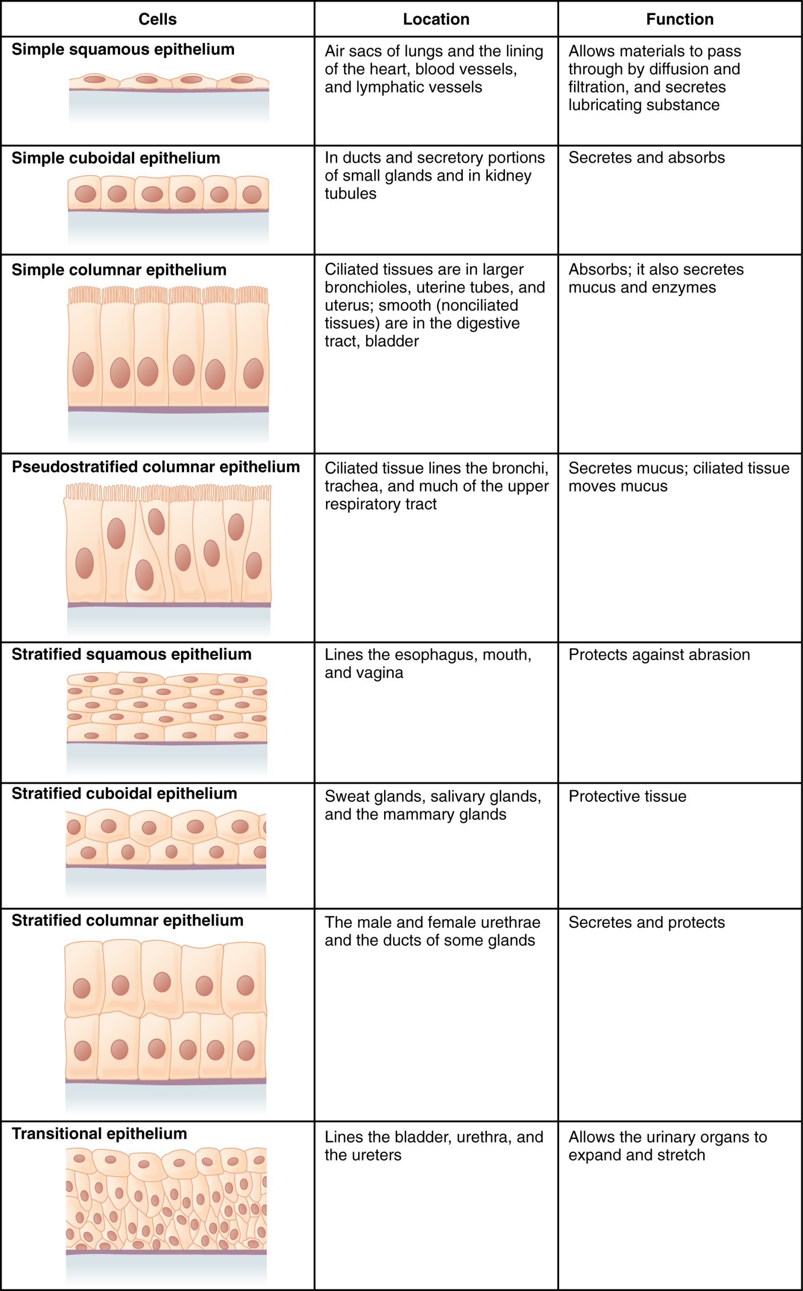
An example of a simple squamous epithelium is the endothelium that lines the vessels of the lymphatic or lymphoid system and blood vessels. Simple cuboidal epithelia are active in the secretion and absorption of molecules. These epithelia are found in the ducts of glands and in the lining of kidney tubules. Simple columnar epithelia also secrete and absorb molecules. These epithelia form in the some sections of the digestive system and in parts of the female reproductive tract. Ciliated columnar epithelia are composed of simple columnar epithelial cells with cilia (small hair like structures). These are present in the lining of the fallopian tubes (a part the female reproductive system) and parts of the respiratory system, where the cila help to remove small particles. Pseudostratified columnar epithelium is a type of epithelium that appears to be stratified but instead consists of a single layer of irregularly shaped and differently sized columnar cells. In pseudostratified epithelium, nuclei of neighboring cells appear at different levels rather than clustered in the basal end. The arrangement gives the appearance of stratification; but in fact all the cells are in contact with the basal lamina, although some do not reach the apical surface. Pseudostratified columnar epithelium is found in the respiratory tract, where some of these cells have cilia.
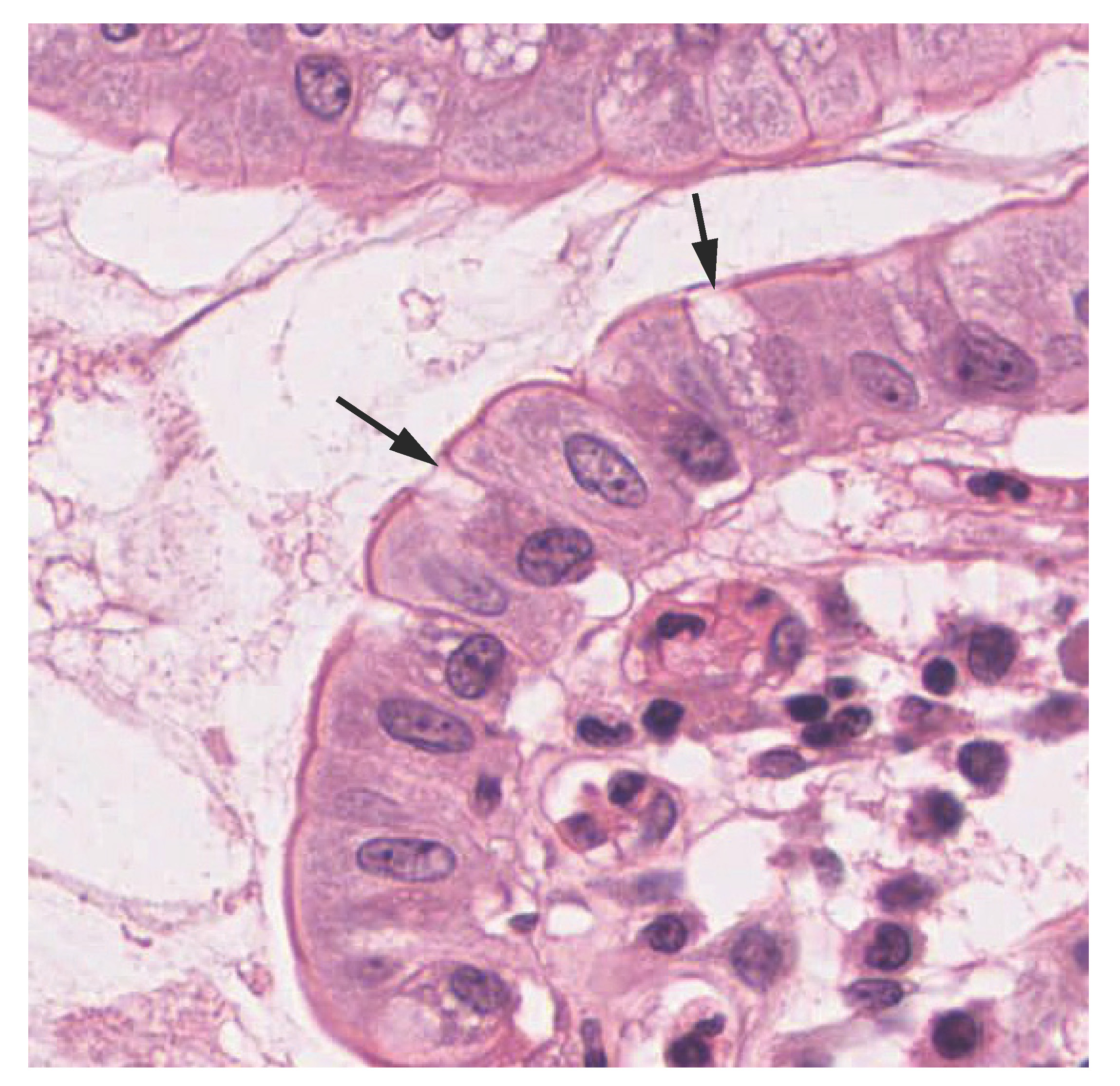
Both simple and pseudostratified columnar epithelia are heterogeneous epithelia because they include additional types of cells interspersed among the epithelial cells. For example, a goblet cell is a mucous-secreting unicellular “gland” interspersed between the columnar epithelial cells of mucous membranes (Figure 5.5).
A stratified epithelium consists of several stacked layers of cells. This epithelium protects against physical and chemical wear and tear. The stratified epithelium is named by the shape of the most apical layer of cells, closest to the free space. Stratified squamous epithelium is the most common type of stratified epithelium in the human body. The apical (top) cells are squamous, whereas the basal layer contains either columnar or cuboidal cells. The top layer may be covered with dead cells filled with keratin. Human skin is an example of this dry, keratinized, stratified squamous epithelium. The lining of the mouth cavity is an example of an unkeratinized, stratified squamous epithelium. Stratified cuboidal epithelium and stratified columnar epithelium can also be found in certain glands and ducts, but are uncommon in the human body. Another kind of stratified epithelium is transitional epithelium, so-called because of the gradual changes in the shapes of the apical cells as the bladder fills with urine. It is found only in the urinary system, specifically the ureters and urinary bladder. When the bladder is empty, this epithelium is convoluted and has cuboidal apical cells with convex, umbrella shaped, apical surfaces. As the bladder fills with urine, this epithelium loses its convolutions and the apical cells transition from cuboidal to squamous. It appears thicker and more multi-layered when the bladder is empty, and more stretched out and less stratified when the bladder is full and distended.
Glandular Epithelium
A gland is a structure made up of one or more cells modified to synthesize and secrete chemical substances. Most glands consist of groups of epithelial cells. A gland can be classified as an endocrine gland, a ductless gland that releases secretions directly into surrounding tissues and fluids (endo- = “inside”), or an exocrine gland who secret their content to the external environment (exo- = “outside”).
Endocrine glands secrete hormones. Hormones are released into the interstitial fluid surrounding the gland cells, diffuse into the bloodstream, and are delivered to cells that have receptors to bind the hormones. The endocrine system is part of a major regulatory system coordinating the regulation and integration of body responses. A few examples of endocrine glands include the anterior pituitary, thymus, adrenal cortex, and gonads.
Exocrine glands release their content to the external environment either directly or through a duct that leads to the epithelial surface. Mucous, sweat, saliva, and breast milk are all examples of secretions from exocrine glands. Secretions into the lumen of the gastrointestinal tract, which is technically outside of the body, are also of the exocrine category. Exocrine glands are classified as either unicellular or multicellular. Unicellular exocrine glands are scattered single cells, such as goblet cells, found in the mucous membranes of the small and large intestine (Figure 5.5).
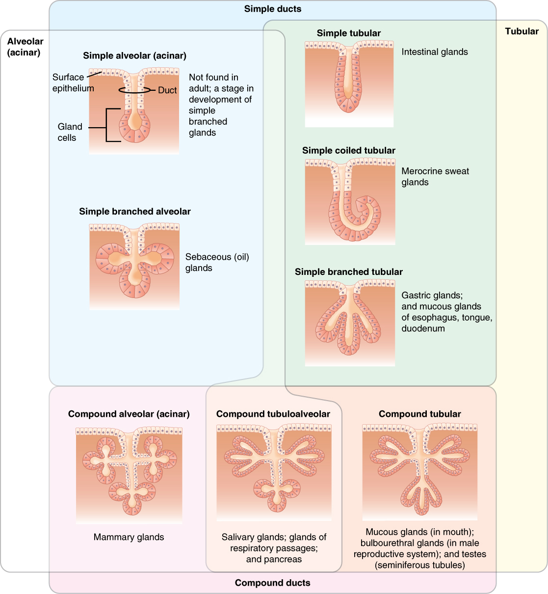
Exocrine glands that release their content through a duct can be classified by their structure and shape of ducts. The ducts can be a single tube, or they can be branched. Figure 5.6 shows a summary of these glands and their classification.
Exocrine glands are also classified by the nature of the substances they release. Serous glands produce a watery, blood-plasma like secretion that is rich in enzymes, such as amylase, an enzyme that breaks down starch and is released in the saliva. Mucous glands secrete a watery to viscous products rich in mucin. Mucin is a so-called glycoprotein that plays an important role in the defense against bacterial and fungal infections as it prevents pathogens to attach to cells. Mixed exocrine glands contain both serous and mucous glands and release both types of secretions.
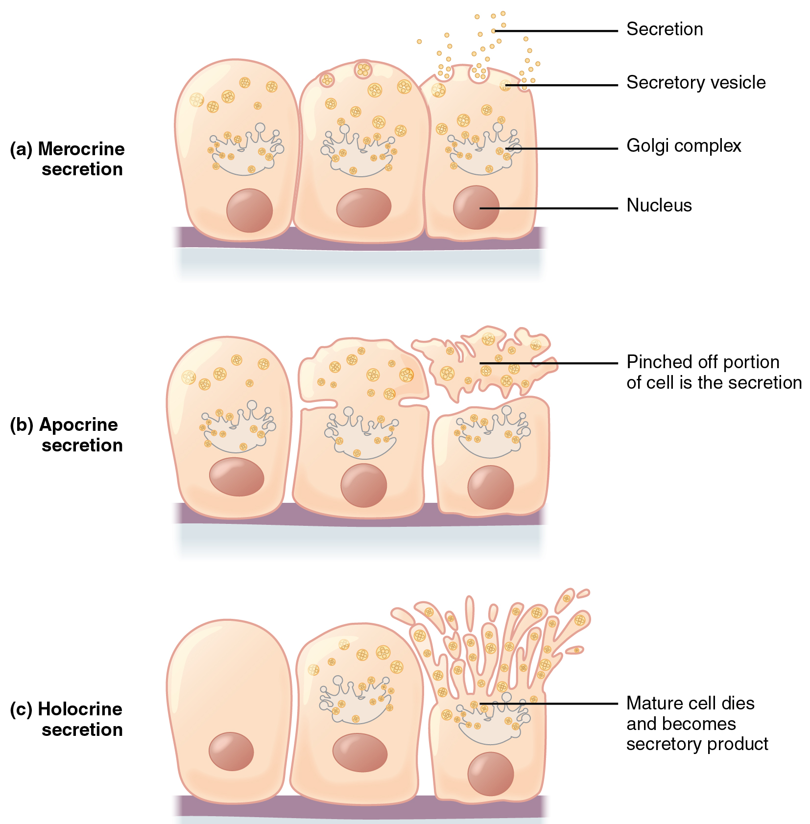
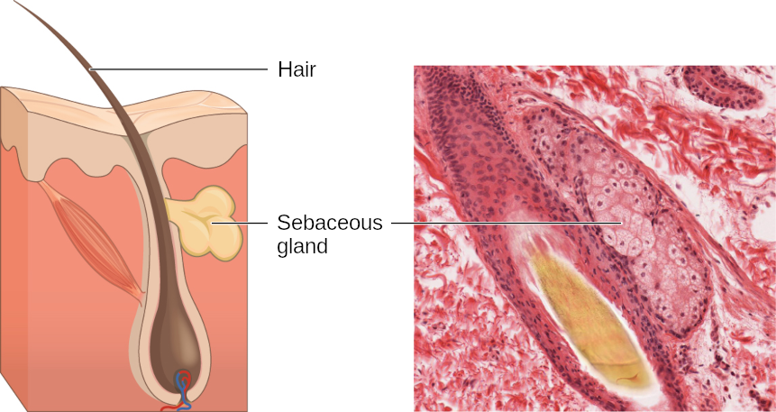
In addition to structure and nature of the secreted substance, exocrine glands can be classified based on how they secrete substances. Figure 5.7 on page depicts the different types of secretion. Merocrine secretion (Figure 5.7a) is the most common type of exocrine secretion. The secretions are enclosed in vesicles that are released by exocytosis. For example, mucin is released by merocrine secretion. Sweat secreted by our skin is an other example. In apocrine secretion (Figure 5.7b) the entire portion of the cell that contains the secretory content is pinched off. An example for glands that release their content by apocrine secretion are sweat glands in our arm pits and crutches. They secrete a fatty substance that local bacteria break down; this causes body odor. Merocrine and apocrine secretion causes little to no damage to the cells. In contrast, in holocrine secretion (Figure 5.7c) the cells accumulate their secretory product and burst to release the content. The gland cells are replaced by new cells from the surrounding tissue. An example are the so-called sebaceous glands that produce the oils on the skin and hair (Figure 5.8).
Key Terms
- apocrine secretion
- release of a substance along with the apical portion of the cell
- basal lamina
- thin extracellular layer that lies underneath epithelial cells and separates them from other tissues
- basement membrane
- in epithelial tissue, a thin layer of fibrous material that anchors the epithelial tissue to the underlying connective tissue; made up of the basal lamina and reticular lamina
- cell junction
- point of cell-to-cell contract that connects one cell to another in a tissue
- epithelium, epithelial tissue
- type of tissue that serves primarily as a covering or lining of body parts, protecting the body; it also functions in absorption, transport, and secretion
- endocrine glands
- groups of cells that release chemical signals into the intercellular fluid to be picked up and transported to their target organs by blood
- endothelium
- tissue that lines vessels of the lymphatic and cardiovascular system, made up of a simple squamous epithelium
- exocrine glands
- group of epithelial cells that secrete substances through ducts that open to the skin or to internal body surfaces that lead toe the exterior of the body
- histology
- microscopic study of tissue architecture, organization, and function
- holocrine secretion
- release of a substance caused by the rupture of a gland cell, which becomes part of the secretion
- merocrine secretion
- release of a substance from a gland via exocytosis
- mucous glands
- group of cells that secrete mucous, a thick, slippery substance that keeps tissues moist and acts as a lubricant.
- pseudostratified columnar epithelium
- tissue that consists of a single layer of irregularly shaped and sized cells that give the appearance of multiple layers; found in ducts of certain glands and the upper respiratory tract
- reticular lamina
- matrix containing collagen and elastin secreted by connective tissue; a component of the basement membrane
- sebaceous glands
- group of cells usually attached to hair follicles that produce a fatty substance
- serous glands
- group of cells within the serous membrane that secretes lubricating substance onto the surface
- simple columnar epithelium
- tissue that consists of a single layer of column-like cells; promotes secretion and absorption in tissues and organs
- simple cuboidal epithelium
- tissue that consists of a single layer of cube-shaped cells; promotes secretion and absorption in ducts and tubules
- simple squamous epithelium
- tissue that consists of a single layer of flat scale-like cells; promotes diffusion and filtration across surface
- stratified columnar epithelium
- tissue that consists of two or more layers of column-like cells, contains glands and is found in some ducts
- stratified cuboidal epithelium
- tissue that consists of two or more layers of cube-shaped cells, found in some ducts
- stratified squamous epithelium
- tissue that consists of multiple layers of cells with the most apical being flat scale-like cells; protects surfaces from abrasion
- tissue
- group of cells that are similar in form and perform related functions
- transitional epithelium
- form of stratified epithelium found in the urinary tract, characterized by an apical layer of cells that change shape in response to the presence of urine
Review Questions
- Question 5.1.1
-
- Question 5.1.2
-
- Question 5.1.3
-

.jpg)